Exploring NYC: The MoMA

One of my favorite exhibits was a collection of modern art from the 1960s. I grew up listening to the music from this decade, and so could appreciate at least some of the cultural references.
When I finished my final exam last Friday, I returned to my room and felt lost. What in the world would I do with 10 days of unstructured time? The answer: have fun! Do things in this grand city that I’ve wanted to do, but simply haven’t had the time (or taken the time) to do. I’ve teamed up with one of my classmates who is also in town this week, and is also casting about for things to do. First on our agenda was to hit The Museum of Modern Art, better known here as The MoMA.
I truly enjoy art, though I don’t pretend to understand all of it. To be honest, much of modern art especially is a mystery to me, though I am fascinated by it. When I read the placards next to the pieces, I can see where the artist is coming from, but often until then … not so much. I think, though, that while part of art may be understanding the thematic and stylistic elements, a significant part is simply experiencing it, the pure visceral nature of the visuals. And that—that I can do.

Vincent van Gogh’s “The Starry Night,” 1889.
Here are some photos I took at The MoMA yesterday. Every piece I’ve captured here intrigued me in some way, though the highlight was probably seeing Vincent van Gogh’s “The Starry Night.” This is undoubtedly one of the most recognizable paintings in existence. How exciting to see it in real life! Most of the other pieces I saw, perhaps with the exception of works by Andy Warhol and Jackson Pollack, were not so familiar to me. But as I said, fascinating nonetheless.

As a writer, how could I NOT love this, Dieter Roth’s “Literature Sausage (Literaturwurst).” According to the exhibit explanation: “Between 1961 and 1970, Roth created about fifty ‘literature sausages.’ To make each sausage Roth followed a traditional recipe, but with one crucial twist: where the recipe called for ground pork, veal, or beef, be substituted a ground-up book or magazine. Roth mixed the ground-up pages with fat, gelatin, water, and spices before stuffing them into sausage casings.” Apparently, he used both materials that he loved and hated, everything from tabloids to Karl Marx. “Roth turned literature into a metaphorical object for intellectual consumption and physical subsistence.” Hm. Well, consider the literature consumed, I suppose.
 I have two photos to share. First, the bottle of brut cava I just bought to put in my refrigerator for tomorrow. Not that I need an excuse to drink bubbly, but this is for a momentous occasion—to mark the end of my first year of medical school. I’m not quite there yet; I’ll be done in 24 hours. With all the hurdles I’ve have had to overcome to get here, finally finishing my first year—not only intact, but truly thriving—is surreal.
 The other photo, and another reason to celebrate: in February of 2017, I will begin my medical “clerkships.” This is where the rubber meets the road, so to speak. They send us out of the classroom and into the hospital to work with real patients (*gulp*). This photo is of my clerkships schedule, which I received yesterday. This will be my life, from February of 2017 to January of 2018: OB/GYN (6 weeks) → Primary care (6 weeks) → Psychiatry (6 weeks) → Surgery (8 weeks) → Anesthesiology (2 weeks) → Open elective time (2 weeks) → Neurology (4 weeks) → Internal Medicine (8 weeks) → Pediatrics (6 weeks).


 Upenuf: It’s an apropos street name for the hilly roads surrounding San Francisco. It’s also an apropos phrase for my life. The months since starting medical school last August have felt very much like an uphill climb. Thankfully, just as I was muttering “upenuf” to myself, we got a week off for spring break. I headed to San Francisco to visit my middle sister, Sarah. What a magical few days! We visited a winery, hiked on the hilly paths of Pacifica, waded in the frigid ocean, walked among the redwoods of Muir Forest, and gazed up (and then down) at the Golden Gate Bridge. We also drank a fair amount of prosecco while eating goat cheese and crackers. I feel rested, relaxed, rejuvenated, and ready for the last push to finish my first year of medical school. Onward … and upward.


 Rembrandt’s facelift. You might be familiar with this famous painting, called the “Anatomy Lesson of Dr. Nicolaes Tulp.” Painted in 1632, it depicts the rare (for the 17th century) event of an autopsy. According to Wikipedia, these were social events with an admission fee. You’re likely not, however, familiar with the photoshopped version of the Rembrandt masterpiece, seen below:
 I looked it up. And according (again) to Wikipedia, it can take a couple forms. In animals, it often involves inserting a tube into the esophagus or stomach, therefore allowing anything that has been swallowed to leak out (and not be digested). Humans probably wouldn’t like that, though. So with people, here is what they do (this is still gross): you smell, taste, and chew the food, finally spitting it out rather than swallowing it. Now I know. And so do you.
 To say the least. Also in our cardiology unit, there was a lecture on thrombosis—known more simply as clotting. Aspirin is something many people take to help prevent clots. But when you’ve got a big one already? Bad news. When this slide of a giant (and potentially fatal) clot came up on the big screen, I just had to jot down what the lecturer said: “At this point, aspirin is not going to help you.”
 That’s just gross. We’re almost done with our pulmonology unit now. One thing we talked a lot about was pneumonia. Realize that pneumonia can be caused by a lot of different bugs. When a particular organism called Klebsiella pneumoniae causes pneumonia, the patient may hack up what’s called “currant jelly sputum”—bloody mucus. Don’t ask how I found this picture. But I did. Someone clearly has way too much time on their hands.
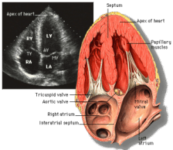
 Friends and colleagues from my former life as a journalist may recognize this notebook as the junior version of the spiral pads we used while employed at the Wednesday Journal, Inc. I discovered it while searching for a notebook that would fit in the pocket of my white coat. This fits the bill without running up the tab—$14.50 for a pack of 12 on Amazon.com. What is a medical history but a specific type of interview? So what better type of notebook to use than one made for a reporter? Just seeing it brings back lots of memories too …