I Heart Ultrasound
by Lorien E. Menhennett
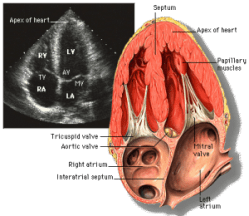
This is an apical four-chamber view of the heart via ultrasound. Key: RV = right ventricle; TV = tricuspid valve; RA = right atrium; LV = left ventricle; AV = aortic valve; MV = mitral valve; LA = left atrium. Image from criticalecho.com.
Even though the focus of the first year of medical school is book-learning, we’re gradually acquiring hands-on skills like taking medical histories and performing a basic physical exam. This week, we got to try something different—cardiac ultrasound. Increasingly used for bedside diagnosis, ultrasound is seen by the Weill Cornell administration as an essential part of our education. It’s also pretty darn cool.
This past Tuesday afternoon, we broke into small groups and rotated through practice exam rooms where we met up with different faculty members and standardized patients. Each group member got a chance to obtain different views of the heart with the ultrasound transducer, including the parasternal long axis, parasternal short axis, apical four chamber, and subxiphoid views.
It was incredible to see the live heart in action, the muscle pumping and the valves fluttering before our eyes, after studying the organ in such great depth for four weeks. The machine we used was pretty nifty too. The screen was not much larger than an iPad. It was mounted standing up, with the ultrasound transducer attached via a cable. Knowing where to put the transducer on the patient’s chest wasn’t so hard—the difficult part was making incremental adjustments to center the image or angle the transducer to better see the chambers or valves. Like everything worthwhile, getting a quality ultrasound image will take practice. But this was a good start.
When it comes to what we’re learning, I like the science. I really do. But it’s clinical activities like this one that remind me of why I’m truly here.
